
The area of overlap of the visual field of one eye with that of the opposite eye is called the binocular field (Figure 14.2). The monocular visual field (Figure 14.1) is determined with one eye covered. Objects within the binocular visual field are visible to each eye, albeit from different angles.

As our eyes are angled slightly toward the nose, the monocular visual fields of the left and right eyes overlap to form the binocular visual field (colored red).

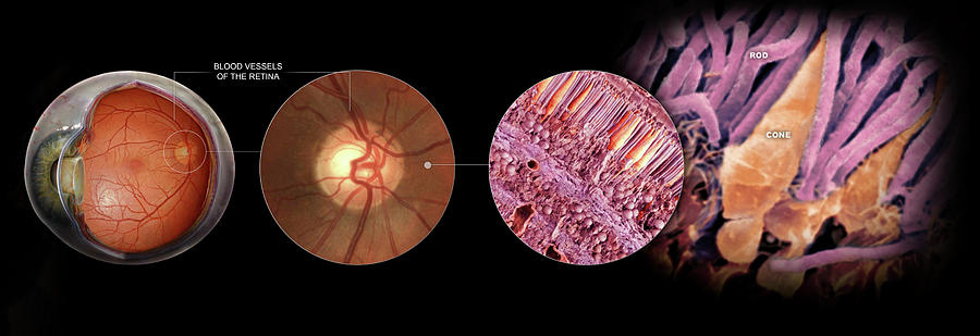
Perimetry testing provides a detailed map of the visual field. As illustrated, the nose prevents the field of the right eye from covering 180 degrees in the horizontal plane. The monocular visual field is the area in space visible to one eye. The visual field is that area in space perceived when the eyes are in a fixed, static position looking straight ahead. The inability to detect objects in specific areas of space (i.e., visual field defects) is often related to neural damage. For example, the ability to detect and identify small objects (i.e., visual acuity) can be affected by disorders in the transparent media of the eye and/or visual nervous system. The condition of the visual system can be determined by examining various aspects of visual sensation. These cells encode different aspects of the visual stimulus, and thus carry independent, parallel, streams of information about stimulus size, color, and movement to the visual thalamus. The information gathered by millions of receptor cells is projected next onto millions of bipolar cells, which, in turn, send projects to retinal ganglion cells. You will learn that the image is first projected onto a flattened sheet of photoreceptor cells that lie on the inner surface of the eye (retina). An important aspect is the regional differences in our visual perception: the central visual field is color-sensitive, has high acuity vision, operates at high levels of illumination whereas the periphery is more sensitive at low levels of illumination, is relatively color insensitive, and has poor visual acuity. The chapter will familiarize you with measures of visual sensation by discussing the basis of form perception, visual acuity, visual field representation, binocular fusion, and depth perception. In this chapter you will learn about how the visual system initiates the processing of external stimuli.


 0 kommentar(er)
